Home /
Expert Answers /
Psychology /
scenario-in-the-nervous-system-afferent-sensory-nerves-conduct-information-from-neurons-innervatin-pa997
(Solved): Scenario: In the nervous system afferent sensory nerves conduct information from neurons innervatin ...
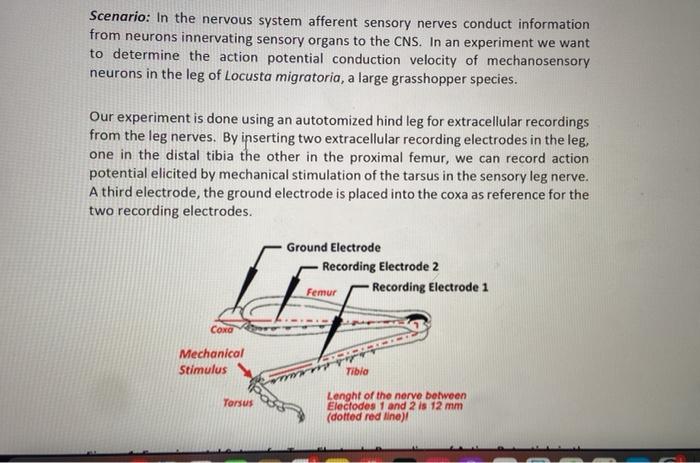
Scenario: In the nervous system afferent sensory nerves conduct information from neurons innervating sensory organs to the CNS. In an experiment we want to determine the action potential conduction velocity of mechanosensory neurons in the leg of Locusta migratoria, a large grasshopper species. Our experiment is done using an autotomized hind leg for extracellular recordings from the leg nerves. By inserting two extracellular recording electrodes in the leg, one in the distal tibia the other in the proximal femur, we can record action potential elicited by mechanical stimulation of the tarsus in the sensory leg nerve. A third electrode, the ground electrode is placed into the coxa as reference for the two recording electrodes. Ground Electrode Recording Electrode 2 Femur Recording Electrode 1 Coxa Mechanical Stimulus wr Tarsus Tibia Lenght of the nerve between Electodes 1 and 2 is 12 mm (dotted red line)!
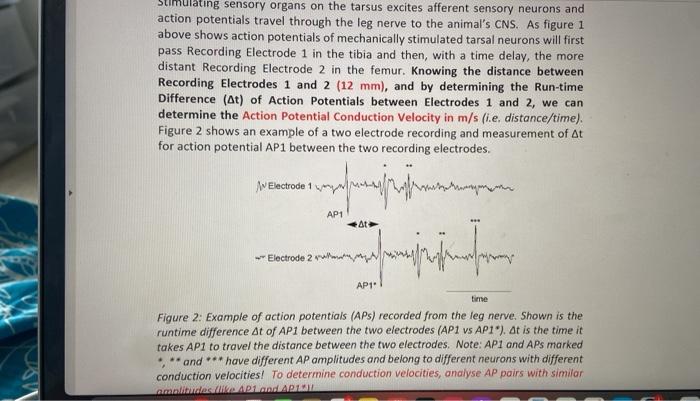
timulating sensory organs on the tarsus excites afferent sensory neurons and action potentials travel through the leg nerve to the animal's CNS. As figure 1 above shows action potentials of mechanically stimulated tarsal neurons will first pass Recording Electrode 1 in the tibia and then, with a time delay, the more distant Recording Electrode 2 in the femur. Knowing the distance between Recording Electrodes 1 and 2 (12 mm), and by determining the Run-time Difference (At) of Action Potentials between Electrodes 1 and 2, we can determine the Action Potential Conduction Velocity in m/s (i.e. distance/time). Figure 2 shows an example of a two electrode recording and measurement of At for action potential AP1 between the two recording electrodes. A Electrode 1 At Jumphrifin form Electrode 2 AP1 I time Figure 2: Example of action potentials (APS) recorded from the leg nerve. Shown is the runtime difference At of AP1 between the two electrodes (AP1 vs AP1). At is the time it takes AP1 to travel the distance between the two electrodes. Note: AP1 and APs marked ***and*** have different AP amplitudes and belong to different neurons with different conduction velocities! To determine conduction velocities, analyse AP pairs with similar amplitudes (like AP1 and API ?? ????? ??????? AP1
2... x 2/5 124% Y Your Tasks for this Assignment: ? 1) Determine the Action Potential Conduction Velocity in m/s from recorded data (data file posted on Moodle)! Use the average of at least three different At measurements from action potential pairings. For averaging conduction velocities pick action potential pairs with similar amplitudes to ensure their comparability. (9 points) ? II) Answer and Explain the following Questions! ? lla) In the extracellular recording from the grasshopper leg we can observe and identify action potentials with very different amplitudes in both traces. What is the reason for different AP amplitudes if APs are All-or- Nothing Responses? Explain this observation! (4 points) ? Ilb) Most of the action potentials are recorded by both electrodes and show up in both recording traces, after all this allows us to calculate their conduction velocities. Provide a reasonable explanation for the situation that action potential *** shown in Figure 2 is only recoded by Electrode 2 in the Femur but not by Electrode 1 in the Tibia! What could be a reason for this observation? Explain your answer! (4 points) D 0 A
• Ilc) Our recording was made at a temperature of 20 °C! What do you expect how the AP conduction velocity will change if this experiment would be repeated at temperatures of (a) 10 °C and (b) 30 °C? Explain! (4 points)
Expert Answer
IIa) An action potential has a similar span and sufficiency like clockwork. Expanded improvement force, then again, produces an expansion in the recurrence of action potentials. The adequacy and span of an action potential don't diminish or reduce as