Home /
Expert Answers /
Biology /
problem-6-3-2-tags-actin-electron-microscopy-mutant-analysis-dyn-pa838
(Solved): Problem 6.3.2 (tags: \#actin, \#electron microscopy, \#mutant analysis, \#dyn ...

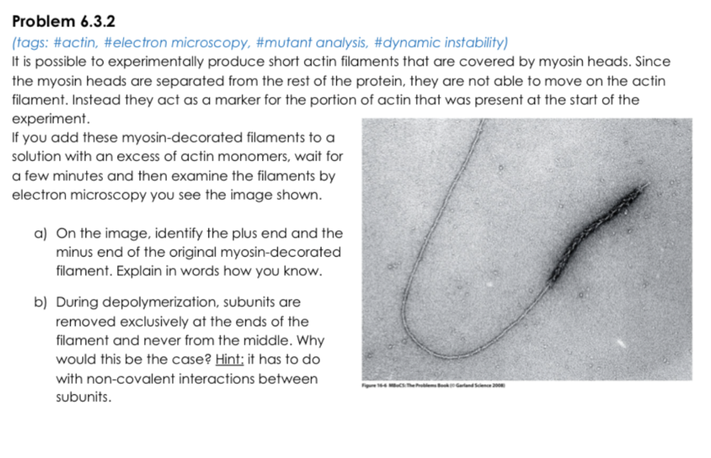
Problem 6.3.2 (tags: \#actin, \#electron microscopy, \#mutant analysis, \#dynamic instability) It is possible to experimentally produce short actin filaments that are covered by myosin heads. Since the myosin heads are separated from the rest of the protein, they are not able to move on the actin filament. Instead they act as a marker for the portion of actin that was present at the start of the experiment. If you add these myosin-decorated filaments to a solution with an excess of actin monomers, wait for a few minutes and then examine the filaments by electron microscopy you see the image shown. a) On the image, identify the plus end and the minus end of the original myosin-decorated filament. Explain in words how you know. b) During depolymerization, subunits are removed exclusively at the ends of the filament and never from the middle. Why would this be the case? Hint: it has to do with non-covalent interactions between subunits.
Expert Answer
The first molecular motor, myosin, is a pro