Home /
Expert Answers /
Biology /
76-laboratory-manual-for-human-biology-activity-3-observing-slides-of-human-skin-materials-for-this-pa343
(Solved): 76 Laboratory Manual for Human Biology ACTIVITY 3 Observing Slides of Human Skin Materials for This ...
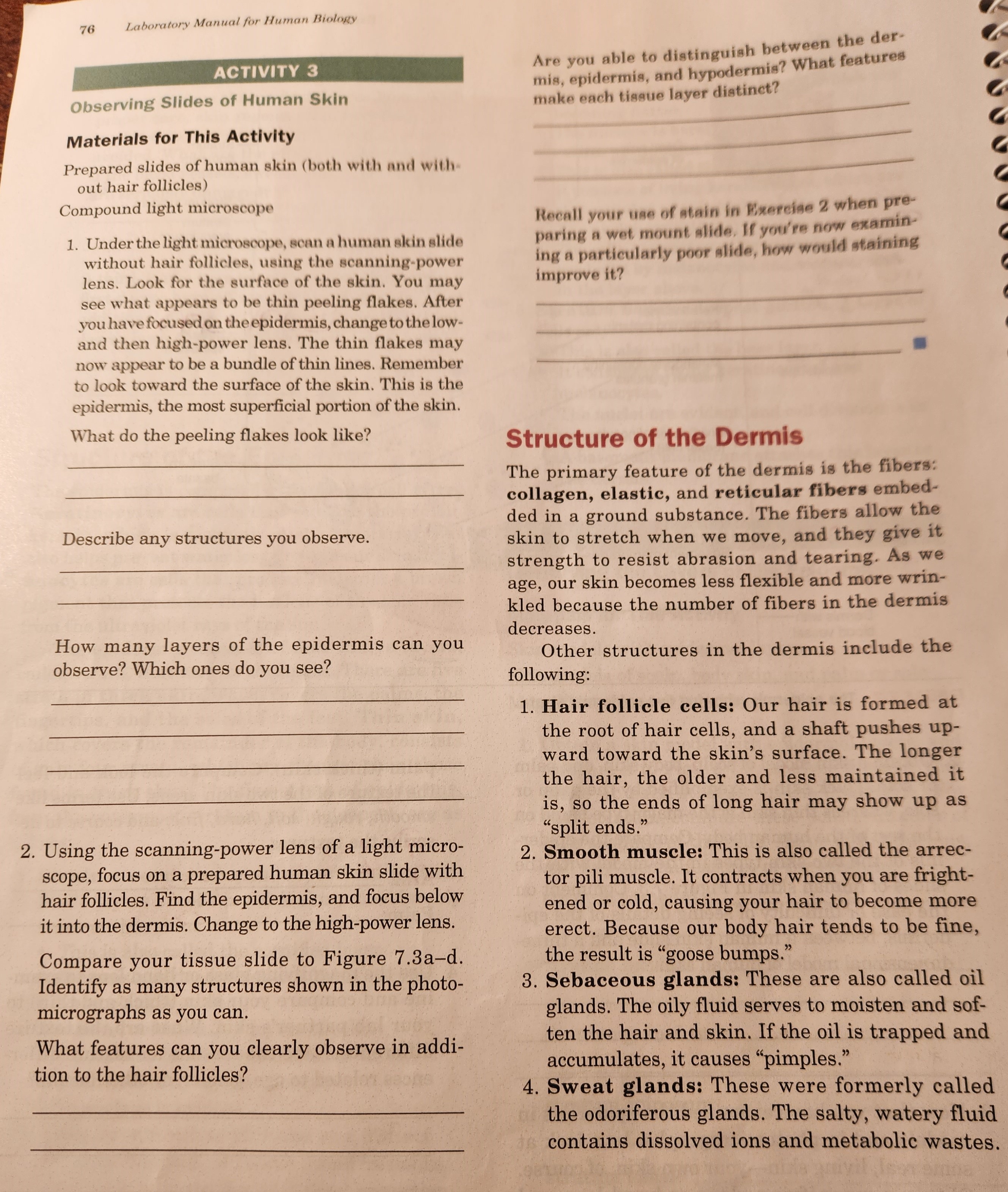
76 Laboratory Manual for Human Biology ACTIVITY 3 Observing Slides of Human Skin Materials for This Activity Prepared slides of human skin (both with and without hair follicles) Compound light microscope 1. Under the light microscope, scan a human skin slide without hair follicles, using the scanning-power lens. Look for the surface of the skin. You may see what appears to be thin peeling flakes. After you have focused on the epidermis, change to the lowand then high-power lens. The thin flakes may now appear to be a bundle of thin lines. Remember to look toward the surface of the skin. This is the epidermis, the most superficial portion of the skin. What do the peeling flakes look like? Describe any structures you observe. How many layers of the epidermis can you observe? Which ones do you see? 2. Using the scanning-power lens of a light microscope, focus on a prepared human skin slide with hair follicles. Find the epidermis, and focus below it into the dermis. Change to the high-power lens. Compare your tissue slide to Figure . Identify as many structures shown in the photomicrographs as you can. What features can you clearly observe in addition to the hair follicles? Are you able to distinguish between the dermis, epidermis, and hypodermis? What features make each tissue layer diatinct? Recall your use of atain in Eirercise 2 when preparing a wet mount alide. If you're now examining a particularly poor slide, how would staining improve it? Structure of the Dermis The primary feature of the dermis is the fibers: collagen, elastic, and reticular fibers embedded in a ground substance. The fibers allow the skin to stretch when we move, and they give it strength to resist abrasion and tearing. As we age, our skin becomes less flexible and more wrinkled because the number of fibers in the dermis decreases. Other structures in the dermis include the following: 1. Hair follicle cells: Our hair is formed at the root of hair cells, and a shaft pushes upward toward the skin's surface. The longer the hair, the older and less maintained it is, so the ends of long hair may show up as "split ends." 2. Smooth muscle: This is also called the arrector pili muscle. It contracts when you are frightened or cold, causing your hair to become more erect. Because our body hair tends to be fine, the result is "goose bumps." 3. Sebaceous glands: These are also called oil glands. The oily fluid serves to moisten and soften the hair and skin. If the oil is trapped and accumulates, it causes "pimples." 4. Sweat glands: These were formerly called the odoriferous glands. The salty, watery fluid contains dissolved ions and metabolic wastes.